
TL;DR
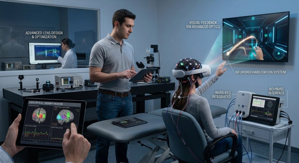
An optical brain-computer interface uses light, rather than electrical signals alone, to read, decode, or influence neural activity. The two main routes are non-invasive methods such as fNIRS and more experimental approaches built on optogenetics and optical imaging. Research is moving quickly, but most advanced methods are still in the laboratory or early clinical stages, and their success depends heavily on precision optical hardware.
Quick Answer
An optical brain-computer interface is a system that uses light to exchange information between the brain and an external device. Optical approaches can offer cell-type specificity, low crosstalk, and scalable channels with high spatial and temporal resolution. The best-known non-invasive method is functional near-infrared spectroscopy (fNIRS), which estimates brain activity from changes in light absorption. More advanced methods use light-sensitive proteins and imaging to record from and stimulate specific neurons.
Introduction
Brain-computer interfaces (BCIs) create a direct communication path between the nervous system and a machine. For decades, most of them have relied on electrodes, which are effective but come with trade-offs in precision, biological compatibility, and the ability to target specific cell types.
Light offers a different path. The optical brain-computer interface is built around photons as the information carrier, and that opens new options for both measuring and influencing neural activity. Instead of only listening to electrical signals, an optical system can watch blood oxygenation, image fluorescent indicators, or deliver pulses of light that switch selected neurons on and off.
For engineers, clinicians, researchers, and product developers, understanding this field is becoming increasingly useful. The Chinese term 光学脑机接口 now appears in recent academic reviews, and the same ideas are discussed worldwide under the English name.
This guide explains what an optical brain-computer interface is, how the main technologies work, where they may be applied, and what challenges remain. It also looks at the optical hardware, from lenses and filters to coatings and micro-displays, that makes these systems possible.
What Is an Optical Brain-Computer Interface?
An optical brain-computer interface, often shortened to OBCI or optical BCI, is a technology that uses optical methods and neuroengineering to enable information exchange between the brain and external devices. It sits at the meeting point of neuroscience, photonics, electronics, and computing.
A helpful way to think about it is as a loop with three stages. Light can be used to read neural activity. A computer then decodes what that activity means. Finally, light can be used to write information back by modulating neurons. Recent reviews describe optical methods as gradually covering this entire read, decode, and write chain, which supports the idea of closed-loop systems that respond to the brain in real time.
Not every optical brain-computer interface covers all three stages. Many current systems only read, as with non-invasive fNIRS headsets. Others focus mainly on stimulation. The most ambitious designs aim to combine both in a single closed loop.
Key Terms You Will Meet
A few terms appear again and again in this field. fNIRS stands for functional near-infrared spectroscopy. Optogenetics refers to the use of light to control genetically modified, light-sensitive cells. Opsins are the light-sensitive proteins involved. Calcium and voltage indicators are molecules that change their fluorescence when a neuron becomes active. Understanding these four ideas makes most of the literature much easier to follow.
Why Use Light? The Case for Optical Approaches
Electrical interfaces are mature and fast, so it is fair to ask why researchers look to light at all. The answer lies in what light can do that electrodes find difficult.
First, light can be highly specific. With the right tools, researchers can target particular types of cells rather than stimulating everything near an electrode. Second, optical signals can show activity across a wide field of view, which allows many neurons to be observed together. Third, optical methods can offer low crosstalk, meaning the stimulation and the recording interfere with each other less than they often do with electrical methods. Finally, an optical brain-computer interface can support multiple measurements at once, such as activity, blood flow, and cell-level signals.
None of this makes light a replacement for electricity. It makes optical methods a complement, strongest where cell-level precision or non-invasive access matters most.
How Does an Optical Brain-Computer Interface Work?
The answer depends on which technology is used. Three routes dominate current research and early clinical work.
Functional Near-Infrared Spectroscopy (fNIRS)
fNIRS is the most mature optical approach and is non-invasive. It sends near-infrared light through the scalp and skull, then measures how much light returns to a detector. Because active brain regions change local blood oxygenation, the amount of light absorbed shifts too. Those shifts can be used to estimate which areas are working harder.
Devices based on fNIRS have already been developed and applied, and this route is widely regarded as a main direction for non-invasive optical brain-computer interface medical devices. Compared with some other brain-monitoring methods, fNIRS is portable and generally more tolerant of body movement and electrical interference. That makes it practical outside a specialist laboratory, for instance at the bedside or in rehabilitation settings.
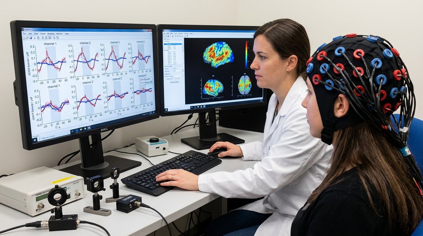
The trade-off is depth and speed. Because the signal follows blood flow, it responds more slowly than electrical activity, and because light scatters in tissue, it mainly reaches the outer layers of the brain.
Optogenetics
Optogenetics takes a very different approach. Neurons are made sensitive to light, typically through genetic techniques, so that specific cells can be activated or silenced using precisely timed pulses of light. This allows a high degree of control over which cells are targeted and when.
Reviews note that optogenetic interfaces address several shortcomings of electrical stimulation, including biocompatibility, stimulation accuracy, and cell-type specificity. That is why an optogenetic optical brain-computer interface has become a hotspot in neural engineering. Researchers have explored it for closed-loop control of brain activity, artificial sensory feedback, and even communication channels between brains in animal experiments.
It is important to be realistic, however. Most of this work has been demonstrated in animals. Because it involves genetic modification, moving it into routine human use raises scientific, safety, and ethical questions that are still being worked through.
Optical Calcium and Voltage Imaging
The third route uses indicators that change brightness when neurons fire. Genetically encoded calcium and voltage indicators allow neural activity to be imaged at cellular and even subcellular resolution. Meanwhile, light-sensitive opsins allow precise control of stimulation or inhibition. Used together, these tools support both recording and stimulation with light.
This is where hardware becomes especially interesting. To image activity deep in tissue, some researchers have developed miniature probes that place light sources, filters, and photodetectors right next to neurons. Bringing the optics close to the cells helps overcome the scattering problem that limits non-invasive methods.
How Signals Travel in a Typical Optical BCI
To make the concept concrete, consider a simplified fNIRS-style pathway. A light source emits near-infrared light into the scalp. Some of that light passes through tissue, is partly absorbed by blood, and returns to a nearby detector. The detector converts the returning light into an electrical signal, which is amplified and digitized. Software then filters out noise from movement and ambient light, estimates changes in oxygenation, and passes the result to a decoding stage.
Depending on the application, that output might be shown to a clinician, used for rehabilitation feedback, or mapped to a command for an external device. Each step introduces its own possible errors, which is why the quality of the optics at the start of the chain matters so much. An optical brain-computer interface can only decode what its sensors manage to capture cleanly.
Optical vs Electrical Brain-Computer Interfaces
Neither approach is simply better. They suit different goals, and many future systems may combine both.
Electrical BCIs are well established and offer fast signal capture. They have a long research record and a clearer path in some clinical settings. An optical brain-computer interface, on the other hand, shows distinct advantages in spatial resolution, cell-type specificity, and the ability to monitor several parameters at the same time.
The trade-off is complexity. Optical methods can require specialized light sources, sensitive detectors, careful alignment, and, in the case of optogenetics, genetic modification. These requirements add scientific and regulatory hurdles.
In practical terms, the comparison looks like this. For a portable, non-invasive system today, fNIRS is the more accessible choice. For high-precision research and possible future closed-loop therapies, optogenetic and imaging-based interfaces are the more ambitious path. Hybrid designs that combine small light sources with electrodes on the same flexible probe are also being explored.
Invasive vs Non-Invasive Optical Systems
Another useful distinction is how close the hardware gets to the brain.
Non-invasive systems sit on the scalp. They are safer and easier to use, which makes them attractive for rehabilitation, assessment, and monitoring. Their limits are depth and resolution, because light has to pass through skin, bone, and tissue before reaching a detector.
Invasive or minimally invasive systems place optical elements closer to or inside neural tissue. They can reach deeper regions and achieve much finer resolution, but they carry surgical risk and face stricter regulatory scrutiny. Most of the high-precision optogenetic and imaging work currently falls into this category and remains in the research stage.
Choosing between them is rarely about technology alone. It depends on the clinical or scientific goal, the acceptable level of risk, and the evidence available.
Potential Applications of Optical Brain-Computer Interface Technology
Non-invasive forms of the optical brain-computer interface are already being explored in clinical and research settings. Functional near-infrared spectroscopy is used in stroke-related motor rehabilitation training, quantitative assessment of brain function in psychiatric conditions, early evaluation of dementia and cognitive impairment, and monitoring of consciousness level. These uses benefit from the portability and safety profile of non-invasive light-based sensing.
Researchers are also investigating how an optical brain-computer interface could support neuroscience research itself, giving scientists a cleaner view of how neural circuits behave in real time. In animal studies, optical methods have been used to test closed-loop control of brain activity and to provide artificial sensory feedback.
Looking further ahead, integrated and miniaturized optical interfaces may support more advanced therapies and richer human-machine interaction. These remain largely research-stage, so claims about near-term consumer products should be treated with caution. Responsible coverage of the field separates what has been demonstrated in people from what has only been shown in the lab.
Common Misconceptions
A few misunderstandings come up often. The first is that an optical brain-computer interface can “read thoughts”. Current systems do not do this. They detect patterns of activity or blood-flow changes linked to specific tasks or states, and interpreting them requires careful calibration and context.
The second is that every optical brain-computer interface involves surgery or genetic changes. In reality, non-invasive fNIRS systems need neither. Genetic modification is specific to optogenetic approaches, which are mainly confined to research.
The third is that light-based systems will simply replace electrodes. A more realistic expectation is a mix of technologies, with each used where it performs best. Treating the optical brain-computer interface as a complement rather than a rival gives a clearer view of where the field is heading.
The Role of Optical Components in an Optical Brain-Computer Interface
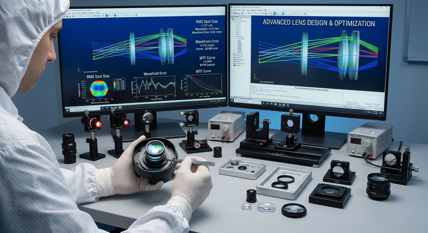
Behind every system sits a chain of optical hardware, and this is where neuroscience meets precision manufacturing. The quality of that hardware shapes what the whole system can detect.
Light Sources and Detectors
Light sources such as LEDs, micro-LEDs, and lasers must deliver stable, well-controlled illumination at the right wavelengths. In fNIRS, near-infrared wavelengths are used because they pass through tissue better than visible light. Detectors then have to capture very weak returning signals, so sensitivity and low noise matter a great deal.
Lenses, Windows, and Prisms
Lenses collect and focus light, whether it is being delivered to tissue or gathered from it. Optical windows protect sensitive sensors and light sources while allowing light to pass. In compact devices, prisms and other folding elements can help fit a useful optical path into a small, wearable form factor. For headsets and head-mounted devices, comfort and weight add further limits on how large the optics can be.
Filters and Coatings
Filters are central to fluorescence-based methods. They separate the wavelength of interest, such as the glow from an activity indicator, from background and excitation light. A weak signal can easily be lost without good filtering. Anti-reflective and other thin-film coatings reduce unwanted reflections and improve transmission, which can make the difference when the signal being measured is already faint.
Micro-Displays and Micro-Lighting
Research groups have also integrated tiny light-emitting structures onto chips for neural stimulation and imaging. Some designs combine multiple colors of micro-emitters with detectors in very small probes. This mirrors trends in micro-display and micro-lighting technology used in other fields such as AR and VR, which shows how progress in one area of optics can benefit another.
Miniaturization
Miniaturization is one of the most visible trends. Researchers have described needle-like imagers that integrate light sources, filters, and photodetectors in a form smaller than 100 micrometers in width, designed to image neural populations at depth. Developments like this show how closely neurotechnology depends on optical design, thin-film coating, and precision manufacturing. Readers interested in the hardware side may want to explore related topics, such as optical component design, optical filters, and optical coating services.
What to Consider When Developing an Optical Brain-Computer Interface Device
Anyone designing or sourcing parts for an optical brain-computer interface faces a distinct set of practical questions.
Wavelength selection comes first. The chosen wavelength determines how deep light can travel, how it interacts with tissue, and which components will perform well. Optical efficiency follows, because small losses at every surface add up in a system where signals are already weak.
Size, weight, and thermal behavior matter just as much, particularly for wearable or implantable designs. Light sources generate heat, and heat near tissue is a safety consideration. Biocompatibility and sterilization become important for anything that contacts the body.
Finally, manufacturing consistency is critical. Medical and research devices need parts that perform the same way from one batch to the next, backed by proper inspection and documentation. Working with a supplier that offers design support and verified testing can reduce the risk of costly redesigns later. For safety-critical medical applications, always follow the relevant regulations and involve qualified engineers and clinicians.
Challenges and Limitations
The optical brain-computer interface faces real obstacles, and understanding them gives a more balanced picture of the field.
Light Scattering in Tissue
Light scatters as it travels through tissue, which limits how deep non-invasive methods can see and how sharp the signal can be. Placing light sources and detectors close to neurons is one way researchers address photon scattering in deep brain tissue, but doing so usually means moving toward more invasive designs.
Clinical Translation
Clinical translation is another hurdle. Optogenetic approaches involve genetic techniques that raise safety and ethical questions. Any medical device also needs rigorous testing and regulatory review before it can be used in patients.
Testing and Standards
Researchers have highlighted the need for suitable testing methods and regulatory approaches for this emerging class of devices. Because an optical brain-computer interface blends optics, electronics, software, and sometimes biology, standard test methods are still developing.
Decoding and Reliability
Signal decoding remains an active research problem. Turning raw optical signals into reliable commands or insights requires robust algorithms, and results from animal studies do not automatically carry over to people. Individual differences in skull thickness, skin, and hair can also affect non-invasive measurements.
Ethical and Safety Considerations
Any technology that interacts with the brain raises questions beyond engineering. Privacy is one of them. Neural data is deeply personal, and its collection, storage, and use need strong safeguards. Informed consent is another, especially where devices may influence brain activity rather than only observe it.
Safety is equally important. Light intensity, exposure time, and heat all need to stay within accepted limits. Developers and clinicians should follow applicable medical-device regulations and guidance from relevant authorities, and readers should treat claims about therapeutic benefit with care until they are backed by solid clinical evidence.
The Future of Optical Brain-Computer Interface Technology
The direction of travel is toward more integrated, smaller, and more capable systems. Reviews point to the potential of closed-loop designs that read, decode, and write neural activity using light, as well as multimodal approaches that combine optical sensing with other measurements.
Progress is likely to come from several directions at once. Better indicators and light-sensitive proteins may improve what can be recorded and controlled. Advances in micro-LEDs, detectors, and on-chip optics may make probes smaller and more efficient. Improved decoding algorithms may turn complex optical signals into more reliable outputs. At the same time, clearer standards for testing and regulation will help promising technologies reach patients more safely.
For the optics industry, this creates interest in precise, low-loss, and compact components, including filters, lenses, windows, and coatings made to tight specifications. The relationship between neuroscience and optical manufacturing is likely to deepen as the field matures.
Conclusion
The optical brain-computer interface represents a promising shift in how the brain can be monitored and, eventually, influenced. Non-invasive fNIRS is already finding practical use in rehabilitation and assessment, while optogenetics and optical imaging point toward higher-precision, closed-loop systems that are still maturing.
The key takeaway is balance. The potential is significant, but so are the technical, clinical, and regulatory challenges. Progress will depend not only on neuroscience but also on dependable optical engineering, from light sources and detectors to filters and coatings.
If you are following or entering this field, start by understanding the differences between the main technologies, be realistic about what has been proven in people, and pay close attention to the quality of the optical hardware. Anyone considering clinical or commercial applications should follow current medical guidance and consult qualified specialists.
Frequently Asked Questions
What is an optical brain-computer interface?
An optical brain-computer interface is a system that uses light to read, decode, or influence brain activity and connect it to an external device. It includes non-invasive methods like fNIRS as well as research-stage approaches such as optogenetics.
What does OBCI stand for?
OBCI stands for optical brain-computer interface. It is sometimes written as optical BCI, and in Chinese as 光学脑机接口.
Is an optical brain-computer interface invasive?
It depends on the method. fNIRS is non-invasive and works through the scalp. Optogenetic and some imaging approaches are more invasive and are mainly used in research settings.
What is the difference between fNIRS and optogenetics?
fNIRS measures changes in light absorption linked to blood oxygenation to estimate brain activity from outside the head. Optogenetics uses light to control specific light-sensitive neurons, usually requiring genetic modification.
Why are optical components important in an optical brain-computer interface?
Lenses, filters, windows, coatings, and light sources control how light is delivered and detected. Their quality directly affects signal clarity and system reliability, so an optical brain-computer interface is only as good as its optical hardware.